
Understanding Traumatic Aortic Injury
A diagnosis of Traumatic Aortic Injury (TAI) can be overwhelming. This guide explains the condition, treatment options, and the road to recovery.
What is the Aorta?
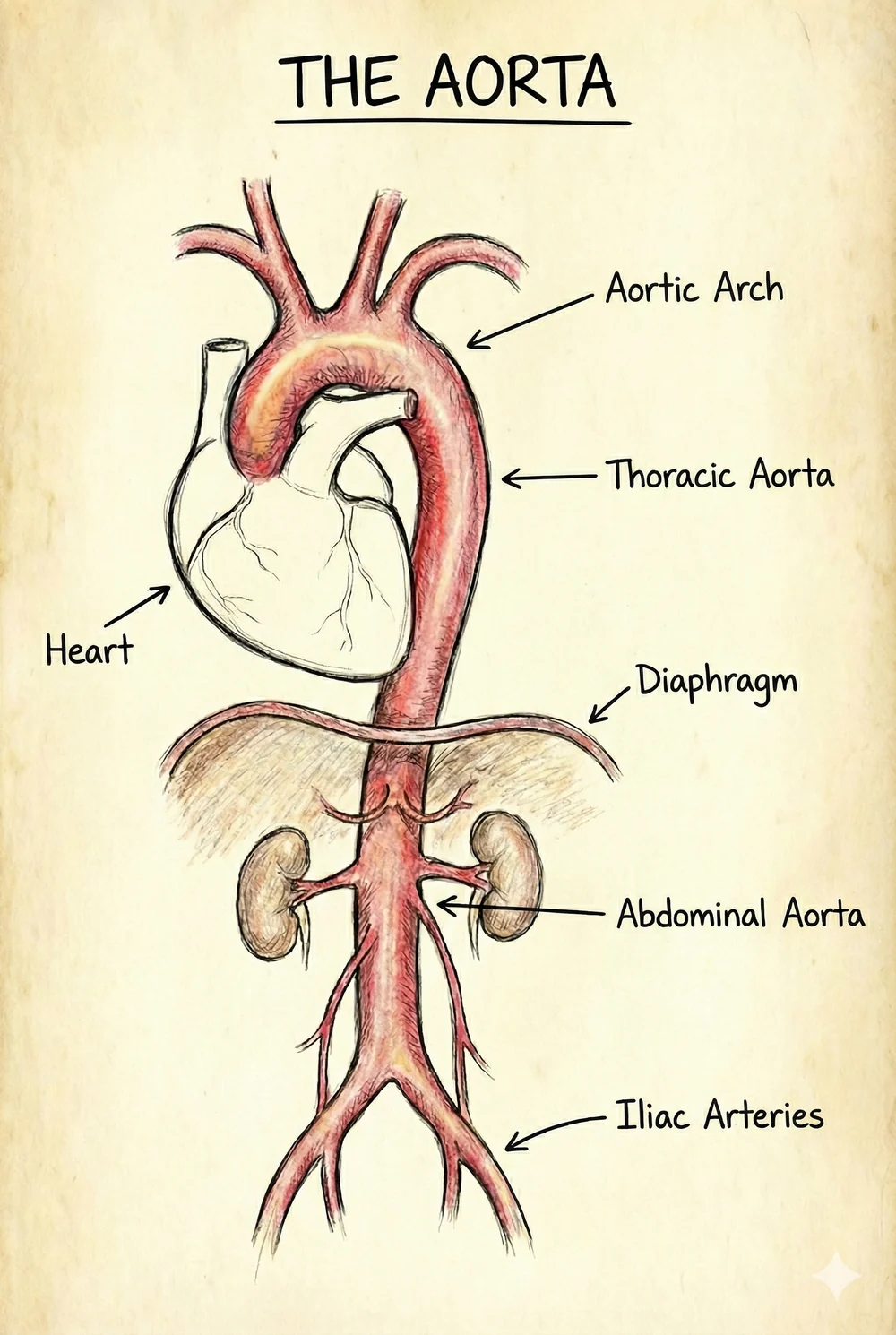
The aorta is the largest blood vessel in your body. It is the main highway that carries oxygen-rich blood from your heart to the rest of your body — your brain, arms, internal organs, and legs.
The section running through the chest — the thoracic aorta — is where traumatic injury most often occurs.
How the Injury Happens
Traumatic Aortic Injury typically occurs during a sudden, high-speed stop, such as a car accident or a fall from a height.
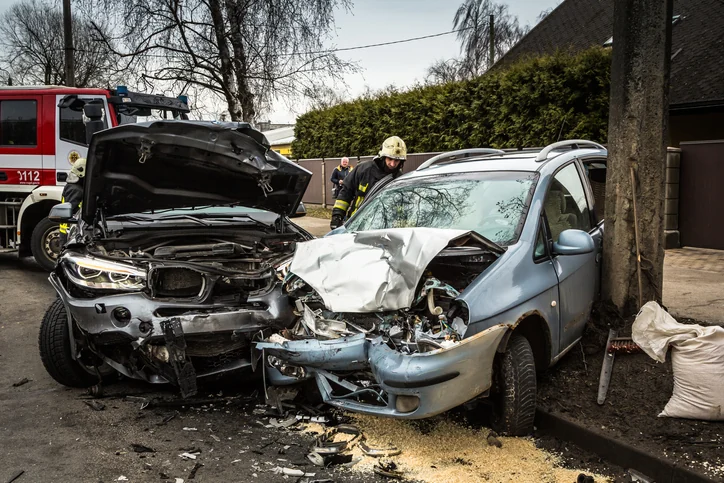
- •Deceleration Force: when the body stops abruptly, the heart and part of the aorta continue forward due to momentum.
- •The Tether Point: a ligament (the ligamentum arteriosum) holds the aorta in place at a spot called the isthmus.
- •The Tear: this shearing force can cause the inner layers of the aortic wall to tear.
Diagnosis & Severity
How is it detected?
Because patients often have other serious injuries from the accident, TAI can be "silent" (no specific symptoms). It is usually diagnosed using a CT Angiogram (CTA), a rapid X-ray scan that uses dye to visualize blood vessels clearly.
The 4 Grades of Injury
Doctors classify the injury into four grades to decide the best treatment.
A small tear on the innermost lining. Treated with medication alone. Routine follow-up scans are not usually needed.
Blood collects within the wall layers. Treated with medication. One follow-up CT scan (CTA) at 1–3 months is recommended to confirm it has resolved.
A bulge forming on the outer wall. Usually repaired with TEVAR. If you are stable, the repair is often planned after the first 24 hours, so other injuries can be treated first.
The wall has burst. Requires emergency repair to stop bleeding.
Symptoms
While often overshadowed by other injuries, conscious patients might report:
- Chest or mid-back pain
- Shortness of breath
- Hoarseness or difficulty swallowing
Note: Absence of pain does not rule out injury.
1. Medical Management
For Grade I, II & Pre-OpAlso known as "Anti-Impulse Therapy." The goal is to lower blood pressure and heart rate to reduce stress on the aortic wall, preventing the tear from getting worse.
Under current guidelines, medical management is the recommended treatment for Grade 1 and Grade 2 injuries — not surgery.
If you also had a head injury: your team may deliberately not lower your blood pressure aggressively. Protecting blood flow to the brain takes priority. This is a considered, guideline-based decision, not an oversight.
Strict BP Control
Crucial for stopping injury progression.
2. TEVAR (Endovascular Repair)
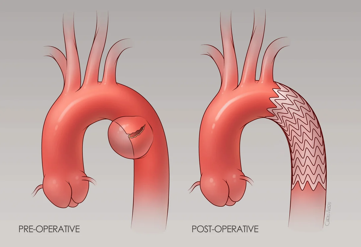
Standard of Care for Grade III & IVThoracic Endovascular Aortic Repair (TEVAR) is a minimally invasive procedure that has largely replaced open chest surgery.
How it works: Surgeons insert a catheter through a small artery in the groin (femoral artery). A collapsed stent graft — a fabric-covered metal tube — is guided up to the aorta.
The repair: Once at the injury site, the stent is deployed. It expands like a spring, pressing against the healthy vessel walls to bridge the tear. Blood then flows through the stent instead of pushing against the injured wall.
Benefits
The Road to Recovery
Phase 1 — Hospital Stay
Most patients spend 24–48 hours in the ICU for strict blood pressure monitoring. If there are no other major injuries, walking usually begins within a day. Pain management is key during this phase.
Phase 2 — Follow-up Imaging
If you had a TEVAR repair, you will have follow-up scans to confirm the stent graft is in position and sealing correctly. There is no single fixed schedule: your team sets one based on your injury, your repair, and your other injuries. Ask your surgeon what your plan is.
Phase 3 — Long Term
TEVAR is designed to be permanent, and long-term follow-up remains important. Managing your blood pressure through diet, exercise, and medication is the most effective way to protect your repair.
A note on recovery
Recovery isn't only physical. Many survivors experience anxiety about their device — this is normal. A support group or a counselor who understands trauma can help during the first year.
Frequently Asked Questions
Most modern thoracic stent grafts are "MRI Conditional," meaning they are generally safe under specific scanning conditions. Always inform your radiology technician and doctor that you have a stent implant before an MRI so they can adjust the machine settings.
It is very unlikely — the amount of metal in a stent graft is minimal. We recommend carrying your device implant card (provided at discharge) in your wallet when traveling, just in case.
Most patients return to normal activities. Heavy lifting and high-impact contact sports may be restricted for several months to allow the aorta to heal. Always consult your surgeon before resuming strenuous exercise.
Call your doctor immediately if you experience sudden chest or back pain, unexplained fever, or any new weakness or numbness in your legs. While rare, these could indicate an issue with the repair and require immediate evaluation.
No. The aorta has no nerve endings that sense touch, so you will not feel the stent inside your chest. Once recovered from the procedure, you should have no physical sensation of the device.
TEVAR devices are designed to be permanent, and long-term data shows they are durable. Follow-up imaging is recommended after TEVAR to confirm the stent stays in position and no "endoleaks" develop. There is no single fixed schedule — your care team will set the one that is right for you.
An endoleak means blood is still flowing into the space between the stent graft and the original aortic wall, usually because the seal is incomplete. Many cause no symptoms and are found only on a surveillance scan. Some resolve on their own; others need treatment — which is why follow-up imaging matters.
Many patients remain on blood pressure medication afterward, because controlled blood pressure reduces stress on the aortic wall and protects the repair. Whether you stay on it, and at what dose, is a decision for your own physician.
This depends less on the aortic repair than on your other injuries, pain levels, and any medication affecting alertness. Most people resume once off strong pain medication and able to move and react normally. Ask your surgeon for clearance first.
Surveillance CT does involve radiation, and it is a reasonable thing to ask about. Current guidelines recommend follow-up imaging after a TEVAR repair but deliberately do not set one fixed schedule for everyone — your team individualizes it, weighing the exposure against the value of confirming your repair is stable. If you have low-grade injuries treated with medication alone, you may need very little imaging, or none. Raise any concerns with your physician.
Download a printable guide
Take this information with you — save it as a PDF or print it for your records.
Medical disclaimer
The information on this page is for educational purposes only and is derived from current medical guidelines. It is not a substitute for professional medical advice, diagnosis, or treatment. Always seek the advice of your physician with any questions regarding a medical condition. If you think you may have a medical emergency, call your doctor or 911 immediately.